
Echo-cardiograms
Atrium medical is now proud to provide echos and cardiology appointments in-house!
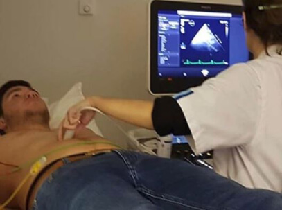
An echo-cardiogram (echo) uses sound waves to create pictures of your heart’s chambers, valves, walls and blood vessels. A probe called a transducer is passed over your chest. The transducer produces sound waves that bounce off your heart and “echo” back to it (hence the name).
What is the difference between an echo and an ECG (electrocardiogram)? An ECG — the squiggly lines most people are familiar with — measures electrical signals and is generally performed during a routine physical exam. On the other hand, the echo provides a direct visual representation of your pumping heart. The echo helps the doctor analyze:
- The size and shape of your heart, and the size, thickness and movement of your heart’s walls.
- How your heart moves.
- The heart’s pumping strength.
- If the heart valves are working correctly.
- If blood is leaking backwards through your heart valves (regurgitation).
- If the heart valves are too narrow (stenosis).
- If there is a tumor or infectious growth around your heart valves
- Problems with the outer lining of your heart (the pericardium).
- Problems with the large blood vessels that enter and leave the heart.
- Blood clots in the chambers of your heart.
- Abnormal holes between the chambers of the heart.
Talk to your provider about cardiac imaging, especially if you have a family history of heart disease, suffer from hypertension, or are prone to other risk factors. A painless, noninvasive and quick test could save your life!